
THIS BLOG HAS BEEN MOVED TO
http://sophiegclarke.tumblr.com/
FOLLOW THERE
xooxox
s
Friday, November 2, 2012
Double Panda Sign
This is an MRI of a brain with Wilson's disease. The disease is a bummer because it causes a lot of psychiatric issues and liver disease. Doctors claim this is because copper accumulates all up in your business but im pretty sure it is because you have TWO PANDAS LIVING IN YOUR BRAIN

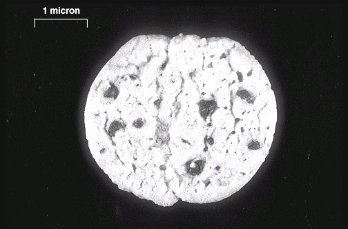
Chocolate Chip Cookies
The cool thing about chocolate chip cookies is that if you eat enough of them, Mr. Von Willebrand factor helps to aggregate all those chocolate chip cookies (that, apparently, are called platelets now but they look just like those cookies you ate) and then you get atherosclerosis and then you die. xoxo. S
One of your cookies:

One of your cookies:
Wednesday, September 5, 2012

Monday, February 13, 2012

Thursday, February 9, 2012
Lymphocyte; Electron Microscope


Some people dont have enough lymphocytes. For example, HIV patients have very few T cells (these are a type of lymphocyte, or white blood cell.) Without these T cells, HIV infected patients are unable to defend themselves against infections that healthy people are unfazed by. The slide above is a white blood cell smear with a lymphocyte smack dab in the middle. It came from the woman embalmed on the pendant below. The pendant is pretty poorly made and some jeweler probably threw it together for a couple of bucks and then sold it to some hipster at an outrageous mark up. I doubt, however, that it was even crappy enough for the hipster. I imagine to "improve it" (so she could claim to her friends it was "vintage"), she hung it from her window and let it rust up in the rain. It is all rather a sad story because the woman (from whom the blood cells above came from and whose face is embalmed on this pendant now owned by a hipster) now looks rather distorted, hole-chested, and horse-like. If you met her in real life, however, you would surely say to yourself "what a beautiful lady.... its too bad really that she smokes so heavily!"... or something to that effect.
Wednesday, February 8, 2012
Brain Sand. H&E


This is a stain of corpora arenacea, otherwise known as brain sand. Brain sand is basically extracellular calcified bodies containing calcium phosphate, magnesium phosphate and carbonate in an organic matrix. Sounds like a lot of BS to me. We have sand.... in our brains? You might as well tell me there are ghosts in my attic!
Tuesday, February 7, 2012
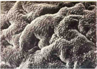
Rugae in the GI tract; Electron Microscope: no stain.

*I used the abbreviated form of the extinct animal because I couldn't even spell the animal correctly enough for this program to autocorrect me.
** I took a little creative license on this photograph. Obviously this dino is climbing a steep rockface (with small high elevation shrubs scattered about), but rocks turn out to be very difficult to draw, so I put this dino in a cave. Apologies if it bothers any of you.
Monday, February 6, 2012
Lipids; H&E


The alien got a little aggressive with his crop circles. He is really gonna freak out the farmers and the idiots who believe in aliens when they wake up in the morning.
Sunday, February 5, 2012
Axons


And just when you thought this blog was getting good, I drew some worms in a traffic jam. Ha. The axons in the slide above are myelinated, which basically means they transmit impulses faster. This is really funny because worms in traffic jams move much more slowly than usual.
Cardiac Muscle; Electron Microscope (No Stain)


Ok so I know this looks like the Midwest (farmland, small ponds, etc) but it is supposed to be much more exotic. Think of the farmland as ripples in a sea of sand dunes and the ponds as merely mirages of wondrous oases. Yes, I KNOW, in reality, this is a slide of cardiac muscle and all of the associated proteins for contraction (myosin, actin, Tropomyosin, Z lines, Intercalated Disks, A bands, H bands, M lines... etc etc etc) but I prefer to look at it and see the view from an airplane window over Tunisia. Spring Break in Tunisia?
Vena Cava Reservoir; H&E


And now, in honor of Downton Abbey (because really, who actually watches the superbowl anyway?) I present you with a woman in a big hat on a Sunday stroll. Unlike most woman her age who have varicose veins (when the things in the slide above are prominent and ugly), she has varicose freckles. It is rather unfortunate for a woman as graceful as she.

Saturday, February 4, 2012
Pericyte; Stain(?)


Pericytes are located outside the endothelium. They help capillaries & venules contract to move blood along. You would need a lot of pericytes to move a bison as big and angry and stubborn as this one along. This bison is very mean. I suspect it is because people have been cutting out bits of him for their gourmet organic grass-fed bison burgers (yum!) with no regard for his comfort or happiness. Fifteen burgers from now, this bison will be nothing. Like the Wicked Witch of the West who left behind only her shoes, this Bison will leave behind only his hair and antlers in a heap on the ground. His only chance for escape is to learn to run like this (below). It is a much faster and more efficient way to get around than pericytes for sure.

Friday, February 3, 2012
Fibrous Cartilage; H&E


These frogs are obviously very sad that their poor friends' legs got chopped off. What idiot, these frogs wonder, would chop off frog legs and not eat them? Waste not want not, my grandmother would say to him. Perhaps the idiot tried them, found them a bit chewy and cartilaginous, and spit them out. It is possible. If you are curious to know what cartilaginous frog legs would look like stained with H&E under a light microscope, I anticipated this and put the slide above. This cartilage is fibrous and contains type I collegen. It is vascular (again, bloody frog legs=yuck) and a row of chondrocytes (cells that lay down cartilage) are stained blue.
Thursday, February 2, 2012
More Bones....H&E


Do mice have bones? I don't know. Small ones if anything. This mouse does, however, have rollerblades and chickenpox. I am not sure if I should feel bad for it or intensely jealous. I used to love those birthday parties at roller rinks. I always won the limbo game and got golden coins to spend at the arcade games where I fared much more poorly.
Wednesday, February 1, 2012
Elastic fibers made from chondroblasts and chondrocytes (Cartilage)


Atlas looks rather uncomfortable. Perhaps it is because the world he is holding is flat and the weight is spread less evenly across his shoulders--or on his mind (the weight of such a fallacy)? My atlas also seems to be chinless. As a barely passing first year medical student, I can say with absolute confidence that I have no idea what anatomical phenomenon this is due to. The collapse of the sternocleidomastoid muscle in the neck perhaps? Instead, I attribute this to years of serious work by me and not enough time using the fun tools (like the spray-painter!) in the computer 'paint' program.
Muscular Vein, Muscular Artery, Nerve, Arteriole; stain?


I can remember 6 obsessions I had as a kid. Card houses, magic tricks, rollerblading, film-making, juggling (soccer balls), and yoyo-ing. I was really only ever good at magic tricks and juggling (see: my recent card house fiasco on Survivor). I did, however, have big dreams to be a professional yoyo-ist. I am not sure what that even is, but I guess had youtube been around those days it would have meant at least a couple hundred thousand views on my best video. The only tricks I ever really mastered were sleeping (is that even considered a trick if you do it with one of those narcoleptic yoyos?) and the one that looked like a swing. Anyways, I was never as adept as this woman above is. She seems also to be able to shake her booty and yoyo at the same time. What an act. What a stupid show off. The slide above is a cross section of a few different types of arteries, veins, and nerves. It really is a beautiful and organized slide. Probably came from this show off woman. Ooo look at me! Yea, we all are.
Tuesday, January 31, 2012
Macrophages in a Connective Tissue Spread: Acid Phosphotase Rxn


Macrophages (shown in the connective tissue stain above) eat things. If skeletal muscle cells were the Survivor contestants, Lacrimal gland cells the bachelor contestants, and Bartholin cells (uh, google it) the Jersey Shore cast members of the human body, Macrophages would be the biggest loser contestants (pre-show and post-show bounce back). They eat and eat and eat. I drew some teeth on this dalmation hoping it would make him look really hungry. Then, I wasnt sure it would come off so I surrounded him with jelly filled doughnuts. The jelly is not seen in the drawing because the donuts haven't been biten into and the drawing is B&W. Anyways, I know all of you 21st century freaks are so damn skeptical of anything B&W (way to ruin it for all of us Hitchcock) that you might suspect my jelly was actually chocolate syrup. So, to maintain the trust we have built this past week, I left the doughnuts unopened. I do promise, though, that there is jelly inside.
Protoplasmic Astrocyte; H&E


Well this is embarrassing. I drew this whole doodle before I realized it actually looked nothing like the astrocyte (thats a nerve cell for all of you people who have lives and dont care about slides, things you cant see with the human eye, chemical staining, and, uh, the mysteries of the universe). So, to compensate, I drew a few dark splotches. I hope they connect the pictures for you. If not, I hope you read this caption before you bothered to look at the photos. Hmm. I guess that, unless you have some sort of weird sort of dyslexia, that is unlikely. Maybe I should have just crossed my fingers and hoped for some sort of The Emperors New Clothes type effect here...
Monday, January 30, 2012
Secondary ossification center; H&E
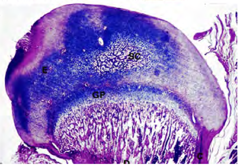

Bones grow longways (interstitial growth) at secondary ossification centers like the one shown on this slide. And, despite what your mothers might have you believe (memories of me crying with milk dripping down my chin as my mother stands over me with a wooden spoon and a tape measure come to mind)-- bones grow quite slowly and even gallons of milk will have no immediate results. In fact, if it was a race, this nice little snail I drew would win every time.
Smooth Muscle. H&E


Exam done. This is smooth muscle. Exam did not go so smoothly. The picture of me lying utterly exhausted in bed is deceptive. My feet are much smaller in real life and I am not smiling right now.
Sunday, January 29, 2012
Epithelium, H&E


I should be studying epithelium right now. Instead, I am dreaming of flying. Spring break to Tunisia?
Saturday, January 28, 2012
Exam Tomorrow. Crap. H&E


I take my joint histology/physiology midterm tomorrow. I have studied 8+ hours a day (ok, well thats a lie, but it makes me seem much more clever than this pathetic little childlike doodle makes me look) since Monday. Fifty-six hours of studying, however, does not necessarily guarantee a passing grade. Likewise, an elephant's size does not guarantee a victory in every Animal Kingdom smackdown (especially against an aligator with teeth as sharp as this one!) Or is this a crocodile? Gosh, I can not even identify my own drawings...
Stereocilia on simple columnar epithelia, H&E


This is a H&E stain of epithelial cells from the penis. I cant remember if it is a section from the ductus deferens or the epididymis, but I doubt even you men know the difference so lets leave it at that. The wispy bits at the top are sterocilia, which aid in secretion and absorption by increasing cell surface area. The picture I drew of the elephants below is almost entirely unrelated. Well, I guess the elephants' trunks are a bit phallic.
Friday, January 27, 2012
Connective Tissue, H&E


This is a spread of connective tissue stained with H&E and Azure. Seen here are some thin elastin fibers and granulitic macrophages. The macrophages help rid the body of worn out cells and debris, and aid in immune response. Although they are usually angels of the immune system, they can sometimes be corrupted by immune system diseases and used for evil. This happens in some cancers, HIV, atherosclerosis, and tuberculosis. It is snowing out so I drew the angel singing Christmas carols. Wikipedia, however, does not say if there is any relationship between macrophages and musical talent. I guess it is possible.
Fetal long bone, ?


Serous Glands, H&E


Shown is a serous gland stained with H&E. These types of glands are found in salivary glands, sweat glands, and tear ducks. On this slide, the gland is secreting hormones in vesicles. This is called apocrine secretion. Although a giraffe's spots look like tiny vesicles, they do not transport anything. I think they are mostly used for camouflage. But, I also read (on wikipedia) that the splotches serve as "thermal windows" where sweat glands, made of serous glands like those shown above, congregate. I kinda think the serous gland slide looks like a giraffe's head with vesicle-googly-eyes. But its late and I am tired.
Subscribe to:
Posts (Atom)